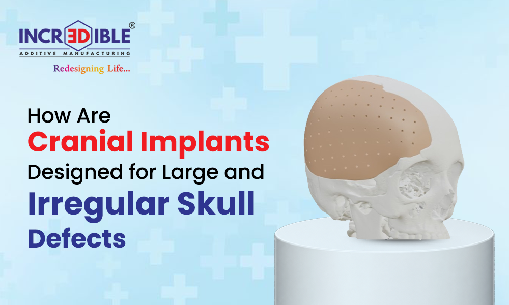
Large or irregular skull defects can be difficult to reconstruct because every patient has a different bone shape, defect boundary, and anatomical requirement. Modern patient-specific cranial implants are designed using medical imaging, 3D modeling, and computer-aided design to closely match the patient’s skull. The process begins with detailed CT imaging and continues through virtual reconstruction, implant design, material selection, and precision manufacturing. This approach helps restore skull contour while supporting the surgeon’s reconstruction plan.
“A cranial implant is not simply shaped to fill a gap; it is designed around the patient’s individual anatomy.”
How Is the Skull Defect Digitally Mapped?
The design process begins with high-resolution CT scans that capture the remaining skull and the boundaries of the defect. The imaging data is converted into a three-dimensional digital model using specialized medical CAD software.
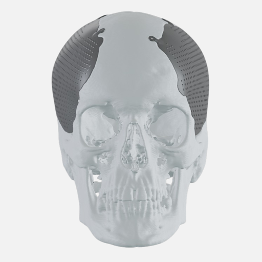
For an irregular or one-sided defect, the intact portion of the skull can help guide reconstruction. Engineers may mirror the corresponding healthy side to create a more natural contour. This digital approach allows custom cranial implants to be designed before fabrication and reviewed as part of the surgical planning process.
CT-based modeling: Converts detailed patient imaging into an accurate three-dimensional representation.
Defect analysis: Defines the size, shape, depth, and boundaries that the implant needs to address.
Symmetry restoration: Uses the unaffected side as a reference where appropriate for reconstructing natural contours.
How Is the Implant Designed for a Large Defect?
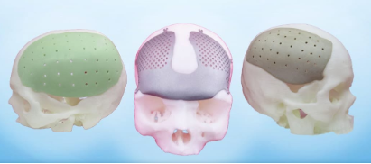
Once the digital skull model is prepared, the implant is shaped to follow the surrounding bone. Designers consider the curvature, implant margins, fixation requirements, and the location of the defect.
For example, a patient with a large unilateral cranial defect following trauma may require an implant that restores the missing contour while providing appropriate fixation points. CAD-based planning allows these features to be incorporated into the design before manufacturing.
Matched contours: Creates implant geometry that follows the surrounding skull surface.
Fixation features: Allows appropriate holes or interfaces for surgical fixation to be incorporated into the design.
Edge optimization: Helps create a controlled transition between the implant and existing bone.
“Digital planning allows the implant geometry to be considered before the physical part reaches the operating room.”
Which Materials Can Be Used for Cranial Implants?
Material selection depends on the clinical requirements, implant design, and surgeon’s preference. Common materials include Ti6Al4V ELI, PEEK, and PMMA, each offering different characteristics.
Titanium alloys provide high mechanical strength and corrosion resistance.
PEEK is radiolucent and has mechanical properties that can resemble bone more closely than metals.
PMMA can be shaped into patient-specific forms and is widely used in reconstructive
The selected material must be suitable for the intended medical application.
How Does 3D Printing Support Cranial Reconstruction?
Once the design is finalized, 3D printed cranial implants can be manufactured using suitable additive manufacturing processes. The digital model guides production, allowing complex contours and patient-specific features to be reproduced accurately.
For instance, a large cranial defect with an uneven boundary may be difficult to reproduce using a standard prefabricated plate. A digitally designed implant can instead follow the specific defect geometry and incorporate the required fixation features.
“The combination of medical imaging, CAD, and additive manufacturing connects the patient’s anatomy directly to the final implant design.”
Key Benefits
Patient-specific design combines medical imaging, digital modeling, material selection, and precision manufacturing to address complex cranial defects with greater anatomical control.
Anatomical fit: Designs are developed around the patient’s skull geometry, helping create an implant that closely follows the existing bone contours.
Improved symmetry: Mirroring suitable areas of the unaffected skull can help recreate a more natural cranial contour in selected cases.
Design flexibility: Digital workflows can accommodate irregular defect boundaries, complex shapes, and customized fixation requirements.
Material options: Titanium, PEEK, and PMMA provide different mechanical, imaging, and handling characteristics for suitable applications.
Surgical planning: A digital implant model allows its geometry and fixation features to be reviewed before manufacturing and surgical placement.
Conclusion
Cranial reconstruction for large and irregular defects requires more than filling the missing area. It involves understanding the patient’s anatomy, accurately defining the defect, designing an appropriate contour, selecting a suitable material, and manufacturing the implant to the approved specifications.
Patient-specific design: Matches the implant to individual skull anatomy.
Digital accuracy: Uses CT data and CAD modeling for detailed planning.
Complex defect support: Accommodates large and irregular skull geometries.
Material flexibility: Supports the use of established implant materials.
Precision manufacturing: Enables digitally designed implants to be produced with complex geometries.
For complex cases involving implants for large skull defects, 3D Medical brings together digital design and advanced manufacturing to support patient-specific cranial reconstruction requirements.